

Imaging Cytometry
Our Amnis ImageStream MarkII Imaging Flow Cytometer combines speed and analysis abilites of flow cytometry with imagery and thus structural and functional insights. Nuclear translocation, colocalization, internalization, immune synapse studies, chemotaxis, morphology/shape change, stem cell differentiation etc. are among the cellular features that can be analyzed.
Our high-end ImageStream offers:
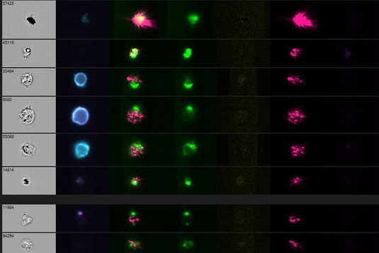
Up to 12 images of every cell:
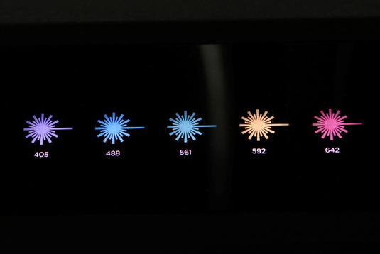
A total of 12 image channels out of 5 lasers (405, 488, 561, 592, 642nm)
can make use of a broad palette of fluorescent markers.
20x, 40x and 60x magnification:
The MultiMag option offer the possibility to obtain cellular localization data of particles ranging from bacteria to large cells.
Extended Depth of Field:
This system upgrade keeps the depth of cell in focus without loss of fluorescence sensitivity, parcticularly used for spot analysis (FISH, acs-specks etc.).
Autosampler for Plates:
Out lates upgrade allowes to run your samples from multiwell plates, enhancing the productivity.
In addition, we provide a workstation - a PC equipped with the IDEAS® software and enough computing capacity to handle the large files resulting from imaging cytometry.
This instrument was financed by the SFB1054, so the usage fees for SFB1054-members are waived.
See below for our instrument configuration described in the official sample preparation guide.
Downloads
- ImageStream_Sample Prep Guide (72 KByte)